A clinical observation published in April 2000 in the Archives of Internal Medicine caught my attention. Dr. Anu Prabhala and his colleagues reported on the treatment of five patients confined to wheelchairs with severe weakness and fatigue. Blood tests revealed that all suffered from severe vitamin D deficiency. The patients received 50,000 IU vitamin D per week and all became mobile within six weeks.1
Dr. Prabhala's research sparked my interest and led to a search for current information on vitamin D, how it works, how much we really need and how we get it. The following is a small part of the important information that I found.
Any discussion of vitamin D must begin with the discoveries of the Canadian-born dentist Weston A. Price. In his masterpiece Nutrition and Physical Degeneration, Dr. Price noted that the diet of isolated, so-called "primitive" peoples contained "at least ten times" the amount of "fat-soluble vitamins" as the standard American diet of his day.2 Dr. Price determined that it was the presence of plentiful amounts of fat-soluble vitamins A and D in the diet, along with calcium, phosphorus and other minerals, that conferred such high immunity to tooth decay and resistance to disease in nonindustrialized population groups.
Today another Canadian researcher, Dr. Reinhold Vieth, argues convincingly that current vitamin D recommendations are woefully inadequate. The recommended dose of 200-400 international units (IU) will prevent rickets in children but does not come close to the optimum amount necessary for vibrant health.3 According to Dr. Vieth, the minimal daily requirement of vitamin D should be in the range of 4,000 IU from all sources, rather than the 200-400 currently suggested, or ten times the Recommended Daily Allowance (RDA). Dr. Vieth's research perfectly matches Dr. Price's observations of sixty years ago!
Vitamin D From Sunlight
Pick up any popular book on vitamins and you will read that ten minutes of daily exposure of the arms and legs to sunlight will supply us with all the vitamin D that we need. Humans do indeed manufacture vitamin D from cholesterol by the action of sunlight on the skin but it is actually very difficult to obtain even a minimal amount of vitamin D with a brief foray into the sunlight.4,5
Ultraviolet (UV) light is divided into 3 bands or wavelength ranges, which are referred to as UV-C, UV-B and UV-A.6 UV-C is the most energetic and shortest of the UV bands. It will burn human skin rapidly in extremely small doses. Fortunately, it is completely absorbed by the ozone layer. However, UV-C is present in some lights. For this reason, fluorescent and halogen and other specialty lights may contribute to skin cancer.
UV-A, known as the "tanning ray," is primarily responsible for darkening the pigment in our skin. Most tanning bulbs have a high UV-A output, with a small percentage of UV-B. UV-A is less energetic than UV-B, so exposure to UV-A will not result in a burn, unless the skin is photosensitive or excessive doses are used. UV-A penetrates more deeply into the skin than UV-B, due to its longer wavelength. Until recently, UV-A was not blocked by sunscreens. It is now considered to be a major contributor to the high incidence of non-melanoma skin cancers.7 Seventy-eight percent of UV-A penetrates glass so windows do not offer protection.
The ultraviolet wavelength that stimulates our bodies to produce vitamin D is UV-B. It is sometimes called the "burning ray" because it is the primary cause of sunburn (erythema).
However, UV-B initiates beneficial responses, stimulating the production of vitamin D that the body uses in many important processes. Although UV-B causes sunburn, it also causes special skin cells called melanocytes to produce melanin, which is protective. UV-B also stimulates the production of Melanocyte
Stimulating Hormone (MSH), an important hormone in weight loss and energy production.8
The reason it is difficult to get adequate vitamin D from sunlight is that while UV-A is present throughout the day, the amount of UV-B present has to do with the angle of the sun's rays. Thus, UV-B is present only during midday hours at higher latitudes, and only with significant intensity in temperate or tropical latitudes. Only 5 percent of the UV-B light range goes through glass and it does not penetrate clouds, smog or fog.
Sun exposure at higher latitudes before 10 am or after 2 pm will cause burning from UV-A before it will supply adequate vitamin D from UV-B. This finding may surprise you, as it did the researchers. It means that sunning must occur between the hours we have been told to avoid. Only sunning between 10 am and 2 pm during summer months (or winter months in southern latitudes) for 20-120 minutes, depending on skin type and color, will form adequate vitamin D before burning occurs.9
It takes about 24 hours for UV-B-stimulated vitamin D to show up as maximum levels of vitamin D in the blood. Cholesterol-containing body oils are critical to this absorption process.10
Because the body needs 30-60 minutes to absorb these vitamin-D-containing oils, it is best to delay showering or bathing for one hour after exposure. The skin oils in which vitamin D is produced can also be removed by chlorine in swimming pools.
The current suggested exposure of hands, face and arms for 10-20 minutes, three times a week, provides only 200-400 IU of vitamin D each time or an average of 100-200 IU per day during the summer months. In order to achieve optimal levels of vitamin D, 85 percent of body surface needs exposure to prime midday sun. (About 100-200 IU of vitamin D is produced for each 5 percent of body surface exposed, we want 4,000 iu.) Light skinned people need 10-20 minutes of exposure while dark skinned people need 90-120 minutes.11
Latitude and altitude determine the intensity of UV light. UV-B is stronger at higher altitudes. Latitudes higher than 30° (both north and south) have insufficient UV-B sunlight two to six months of the year, even at midday.12 Latitudes higher than 40° have insufficient sunlight to achieve optimum levels of D during six to eight months of the year. In much of the US, which is between 30° and 45° latitude, six months or more during each year have insufficient UV-B sunlight to produce optimal D levels. In far northern or southern locations, latitudes 45° and higher, even summer sun is too weak to provide optimum levels of vitamin D.13-15 A simple meter is available to determine UV-B levels where you live.
Vitamin D From Food
What the research on vitamin D tells us is that unless you are a fisherman, farmer, or otherwise outdoors and exposed regularly to sunlight, living in your ancestral latitude (more on this later), you are unlikely to obtain adequate amounts of vitamin D from the sun. Historically the balance of one's daily need was provided by food. Primitive peoples instinctively chose vitamin-D-rich foods including the intestines, organ meats, skin and fat from certain land animals, as well as shellfish, oily fish and insects. Many of these foods are unacceptable to the modern palate and are highly acidic.
For food sources to provide us with D the source must be sunlight exposed. With exposure to UV-B sunlight, vitamin D is produced from fat in the fur, feathers, and skin of animals, birds and reptiles. Carnivores get additional D from the tissues and organs of their prey. Lichen contains vitamin D and may provide a source of vitamin D in the UV-B sunlight-poor northern latitudes.16 Vitamin D content will vary in the organs and tissues of animals, pigs, cows, and sheep, depending on the amount of time spent in UV-B containing sunlight and/or how much D is given as a supplement. Poultry and eggs contain varying amounts of vitamin D obtained from insects, fishmeal, and sunlight containing UV-B or supplements. Fish, unlike mammals, birds and reptiles, do not respond to sunlight and rely on vitamin D found in phytoplankton and other fish. Salmon must feed on phytoplankton and fish in order to obtain and store significant vitamin D in their fat, flesh, skin, and organs. Thus, modern farm-raised salmon, unless artificially supplemented, may be a poor source of this essential nutrient.
Modern diets usually do not provide adequate amounts of vitamin D;17 partly because of the trend to low fat foods and partly because we no longer eat vitamin-D-rich foods like naturally reared poultry and fatty fish such as kippers, and herring.
Often we are advised to consume the egg white while the D is in the yolk or we eat the flesh of the fish avoiding the D containing skin, organs and fat.
Sun avoidance combined with reduction in food sources contribute to escalating D deficiencies. Vegetarian and vegan diets are exceptionally poor or completely lacking in vitamin D predisposing to an absolute need for UV-B sunlight. Using food as one's primary source of D is difficult to impossible.
Vitamin D pH Miracles
Sunlight and vitamin D are critical to all life forms. Standard textbooks state that the principal function of vitamin D is to promote calcium absorption in the gut and calcium transfer across cell membranes, thus contributing to strong bones and a calm, contented nervous system. It is also well recognized that vitamin D aids in the absorption of magnesium, iron and zinc, as well as calcium.
Actually, vitamin D does not in itself promote healthy bone. Vitamin D controls the levels of calcium in the blood. If there is not enough calcium in the diet, then it will be drawn from the bone to help maintain the alkalinity of the blood.
Receptors for vitamin D are found in most of the cells in the body and research during the 1980s suggested that vitamin D contributed to a healthy immune system, promoted muscle strength, regulated the maturation process and contributed to hormone production.
During the last ten years, researchers have made a number of exciting discoveries about vitamin D. They have ascertained, for example, that vitamin D is an antioxidant that is a more effective antioxidant than vitamin E in reducing lipid peroxidation and increasing enzymes that protect against oxidation.19;20
Vitamin D deficiency decreases biosynthesis and release of insulin.21 Glucose intolerance has been inversely associated with the concentration of vitamin D in the blood. Thus, vitamin D may protect against both Type I and Type II diabetes.22
The risk of senile cataract is reduced in persons with optimal levels of D and carotenoids.23
PCOS (Polycystic Ovarian Syndrome) has been corrected by supplementation of D and calcium.24
Vitamin D plays a role in regulation of both the "infectious" immune system and the "inflammatory" immune system.25
Low vitamin D is associated with several autoimmune diseases including multiple sclerosis, Sjogren's Syndrome, rheumatoid arthritis, thyroiditis and Crohn's disease.26;27
Osteoporosis is strongly associated with low vitamin D. Postmenopausal women with osteoporosis respond favorably (and rapidly) to higher levels of D plus calcium and magnesium.28
D deficiency has been mistaken for fibromyalgia, chronic fatigue or peripheral neuropathy.1;28-30
Infertility is associated with low vitamin D.31 Vitamin D supports production of estrogen in men and women.32 PMS has been completely reversed by addition of calcium, magnesium and vitamin D.33 Menstrual migraine is associated with low levels of vitamin D and calcium.81
Breast, prostate, skin and colon cancer have a strong association with low levels of D and lack of sunlight.34-38
Activated vitamin D in the adrenal gland regulates tyrosine hydroxylase, the rate limiting enzyme necessary for the production of dopamine, epinephrine and norepinephrine. Low D may contribute to chronic fatigue and depression.39
Seasonal Affective Disorder has been treated successfully with vitamin D. In a recent study covering 30 days of treatment comparing vitamin D supplementation with two-hour daily use of light boxes, depression completely resolved in the D group but not in the light box group.40
High stress may increase the need for vitamin D or UV-B sunlight and calcium.41
People with Parkinsons and Alzheimers have been found to have lower levels of vitamin D.42;43
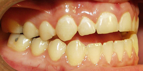
Low levels of D, and perhaps calcium, in a pregnant mother and later in the child may be the contributing cause of "crooked teeth" and myopia. When these conditions are found in succeeding generations it means the genetics require higher levels of one or both nutrients to optimize health.44-47
Behavior and learning disorders respond well to D and/or calcium combined with an adequate diet and trace minerals.48;49
Vitamin D and Heart Disease
Research suggests that low levels of vitamin D may contribute to or be a cause of syndrome X with associated hypertension, obesity, diabetes and heart disease.50 Vitamin D regulates vitamin-D-binding proteins and some calcium-binding proteins, which are responsible for carrying calcium to the "right location" and protecting cells from damage by free calcium.51
Thus, high dietary levels of calcium, when D is insufficient, may contribute to calcification of the arteries, joints, kidney and perhaps even the brain.52-54
Many researchers have postulated that vitamin D deficiency leads to the deposition of calcium in the arteries and hence atherosclerosis, noting that northern countries have higher levels of cardiovascular disease and that more heart attacks occur in winter months.55-56
Scottish researchers found that calcium levels in the hair inversely correlated with arterial calcium-the more calcium or plaque in the arteries, the less calcium in the hair. Ninety percent of men experiencing myocardial infarction had low hair calcium. When vitamin D was administered, the amount of calcium in the beard went up and this rise continued as long as vitamin D was consumed. Almost immediately after stopping supplementation, however, beard calcium fell to pre-supplement levels.27
Administration of dietary vitamin D or UV-B treatment has been shown to lower blood pressure, restore insulin sensitivity and lower cholesterol.58-60
The Battle of the Bulge
Did you ever wonder why some people can eat all they want and not get fat, while others are constantly battling extra pounds? The answer may have to do with vitamin D and calcium status. Sunlight, UV-B, and vitamin D normalize food intake and normalize blood sugar. Weight normalization is associated with higher levels of vitamin D and adequate calcium.61 Obesity is associated with vitamin-D deficiency.62-64 In fact, obese persons have impaired production of UV-B-stimulated D and impaired absorption of food source and supplemental D.65
When the diet lacks calcium, whether from D or calcium deficiency, there is an increase in fatty acid synthase, an enzyme that converts calories into fat. Higher levels of calcium with adequate vitamin D inhibit fatty acid synthase while diets low in calcium increase fatty acid synthase by as much as five-fold.
In one study, genetically obese rats lost 60 percent of their body fat in six weeks on a diet that had moderate calorie reduction but was high in calcium. All rats supplemented with calcium showed increased body temperature indicating a shift from calorie storage to calorie burning (thermogenesis).61
The Right Fats
The assimilation and utilization of vitamin D is influenced by the kinds of fats we consume. Increasing levels of both polyunsaturated and monounsaturated fatty acids in the diet decrease the binding of vitamin D to D-binding proteins.
Saturated fats, the kind found in butter, tallow and coconut oil, do not have this effect. Nor do the omega-3 fats.66
D-binding proteins are key to local and peripheral actions of vitamin D. This is an important consideration as Americans have dramatically increased their intake of polyunsaturated oils (from commercial vegetable oils) and monounsaturated oils (from olive oil and canola oil) and decreased their intake of saturated fats over the past 100 years.
In traditional diets, saturated fats supplied varying amounts of vitamin D. Thus, both reduction of saturated fats and increase of polyunsaturated and monounsaturated fats may contribute to the current widespread D deficiency.
Trans fatty acids, found in margarine and shortenings used in most commercial baked goods, should always be avoided. There is evidence that these fats can interfere with the alkaline buffering systems the body uses to convert vitamin D in the liver.80
Vitamin D Therapy
In my clinical practice, I test for vitamin-D status first. If D is needed, I try to combine sunlight exposure with vitamin D and Vitamin D3 supplementation.
Single, infrequent, intense, skin exposure to UV-B light not only causes sunburn but also suppresses the immune system. On the other hand, frequent low-level exposure normalizes immune function, enhancing NK-cell and T-cell production, reducing abnormal inflammatory responses typical of autoimmune disorders, and reducing occurrences of infectious disease.26;67;68-71
Thus it is important to sunbathe frequently for short periods of time, when UV-B is present, rather than spend long hours in the sun at infrequent intervals. Adequate UV-B exposure and vitamin-D production can be achieved in less time than it takes to cause any redness in the skin. It is never necessary to burn or tan to obtain sufficient vitamin D.
If sunlight is not available in your area because of latitude or season, sunlamps made by Sperti can be used to provide a natural balance of UV-B and UV-A. Used according to instructions, these lamps provide a safe equivalent of sunlight and will not cause burning or even heavy tanning. Tanning beds, on the other hand, are not acceptable as a means of getting your daily dose of vitamin D because they provide high levels of UV-A and very little UV-B.
If you have symptoms of vitamin-D insufficiency or are unable to spend time in the sun, due to season or lifestyle or prior skin cancer, consider adding a supplement of 50,000 IU daily.
Higher levels may be needed but should be recommended and monitored by your health care practitioner after testing serum 25(OH)D.
Supplementation of Vitamin D3 is safe as long as you diet is alkalizing and contains adequate alkalizing minerals such a sodium, calcium, magnesium and potassium.
Adequate calcium and magnesium, as well as other minerals, are critical parts of vitamin D therapy. Without calcium and magnesium in sufficient quantities, vitamin-D supplementation will withdraw calcium from the bone and will allow the uptake of toxic minerals. Do not supplement vitamin D and do not sunbathe unless you are sure you have sufficient calcium and magnesium to meet your daily needs. I suggest a minimum of 1,200-2,400 mg of calcium daily. Research suggests that 1,200-1,500 mg is adequate as a supplement for most adults, both men and women. (Magnesium intake should be half that of calcium.)
Higher amounts of calcium are important for anyone diagnosed with bone loss. Total daily calcium as a supplement may range from 1,500 mg to 2,000 mg depending on current bone status and your body size. Make the effort to split up your daily dose. Do not take all your calcium and magnesium once a day. A higher percentage of the calcium dose is absorbed if delivered in smaller, more frequent amounts.82
Patients on vitamin-D therapy report a wide range of beneficial results including increased energy and strength, resolution of hormonal problems, weight loss, an end to sugar cravings, blood sugar normalization and improvement of nervous system disorders.
A paradoxical transient and non-complicating hypercalciuria (more calcium in the urine) may occur when the program is first initiated. This resolves quickly when adequate calcium and other minerals are consumed. Two other temporary side effects may occur during the first several months of treatment. One is daytime sleepiness after calcium is taken. This usually resolves itself after about one week. The other condition is the reappearance of pain and discomfort at the site of old injuries, a sign of injury remodeling or proper healing, which may take some time to clear up.
Toxicity Issues
Doses used in clinical studies range from as little as 400 IU daily to 10,000-500,000 IU, given either as a single onetime dose or daily, weekly or monthly. Such large doses are given either as a prophylactic or because compliance is considered a problem. There seems to be some evidence that vitamin D works better, without toxicity, when given in lower, more physiologic doses of 2,000-4,000 IU daily rather than as 100,000 IU once a month. However, a single monthly dose of 100,000 IU did replete low levels of vitamin D in adolescents during winter.77
The Many Forms of Vitamin D
There are two types of vitamin D found in nature. Vitamin D2 is formed by the action of UV-B on the plant precursor ergosterol. It is found in plants and in was formerly added to irradiated cows milk. Most milk today contains D3. Vitamin D3 or cholecalciferol is found in animal foods. Both forms of vitamin D have been used successfully to treat rickets and other diseases related to vitamin D insufficiency.
Many consider D3 the preferred vitamin, having more biologic activity. Vitamin D3 as found in food or in human skin always comes with various metabolites or isomers that may have biological benefit.
When humans take in vitamin D from food or sunlight, it is converted first in the liver to the form 25(OH)D and then in the kidney to 1,25(OH)D. These active forms of vitamin D are available by prescription and are given to patients with liver or kidney failure or those with an hereditary metabolic defect in vitamin-D conversion.
Assessing Vitamin D Status
Blood Testing: Currently there are two tests available for physicians to assess vitamin-D status. One is for the somewhat biologically active precursor 25(OH)D and another for 1,25(OH)D, the most active form, which is converted in the kidney and other organs. The latter is often normal in the blood even when the precursor 25(OH)D is low or deficient. The precursor is a better marker of vitamin-D status (or reserves) than the most active 1,25(OH)D form. It is the optimum level of 25(OH)D that is most strongly associated with general good health. (The test values given in this article are for 25(OH)D.) For many years the acceptable level of 25(OH)D has been at least 9 ng/ml (23 nmol/l). Some researchers believe that 20 ng/ml (50 nmol/l) should be the lower acceptable limit72 but Dr. Vieth presents a large amount of data to support his claim that this is far from optimal.3 Optimal levels are certainly at least 32 ng/ml (80 nmol/l) and preferably closer to 40 ng/ml (100 nmol/l).
Salivary pH Testing for calcium sufficiency: A method of assessing ionized calcium levels has been used by Weston Price, DDS and Carl Reich, MD and has confirmation in current research.73 After determining your serum-D status (testing) and undertaking a program of supplementation with vitamin D3, calcium and magnesium, morning salivary pH should read 6.8-7.2. Lower values may indicate insufficient vitamin D (retest), or low levels of calcium in the diet. Look for pH paper with a range of 5.5-8.0 and increments of 0.2 on our website at www.phmiracleliving.com. pH papers with 0.5-degree increments are not sensitive enough to monitor progress.
Sources
* UV-B Meter: Sunsor, Inc. (800) 492-9815 Sunsor
* pH Testing Papers: www.phmiracleliving.com
* Vitamin D3: (888) 880-3055 www.phmiracleliving.com
* Sperti Sunlamps: (800) 544-3757 www.sperti.com
References
1. Prabhala A, Garg R, Dandona P. Severe myopathy associated with vitamin D deficiency in western New York. Arch.Intern.Med. 2000;160:1199-203.
2. Price, Weston A. Characteristics of Primitive and Modernized Dietaries. Nutrition and Physical Degeneration. New Canaan, Connecticut: Keats Publishing, Inc 1989:256-81.
3. Vieth R. Vitamin D supplementation, 25-hydroxyvitamin D concentrations, and safety [see comments]. Am.J.Clin.Nutr. 1999;69:842-56.
4. Glerup H, Mikkelsen K, Poulsen L et al. Commonly recommended daily intake of vitamin D is not sufficient if sunlight exposure is limited. J.Intern.Med. 2000;247:260-8.
5. Glerup H, Eriksen EF. [Vitamin D deficiency. Easy to diagnose, often overlooked (see comments)]. Ugeskr.Laeger 1999;161:2515-21. 6. Diffey BL. Solar ultraviolet radiation effects on biological systems. Phys.Med.Biol. 1991;36:299-328.
7. Moan J, Dahlback A, Setlow RB. Epidemiological support for an hypothesis for melanoma induction indicating a role for UVA radiation. Photochem.Photobiol. 1999;70:243-7.
8. Ranson M, Posen S, Mason RS. Human melanocytes as a target tissue for hormones: in vitro studies with 1 alpha-25, dihydroxyvitamin D3, alpha-melanocyte stimulating hormone, and beta-estradiol. J.Invest Dermatol.1988;91:593-8.
9. Sayre, R. M., Dowdy, J. C., Shepherd, J., Sadig, I., Bager, A., and Kollias, N. Vitamin D Production by Natural and Artificial Sources. 1998. Orlando, Florida, Photo Medical Society Meeting. 3-1-1998. Ref Type: Conference Proceeding
10. Holick MF. The cutaneous photosynthesis of previtamin D3: a unique photoendocrine system. J.Invest Dermatol. 1981;77:51-8.
11. Matsuoka LY, Wortsman J, Haddad JG, Kolm P, Hollis BW. Racial pigmentation and the cutaneous synthesis of vitamin D [see comments]. Arch.Dermatol. 1991;127:536-8.
12. Matsuoka LY, Wortsman J, Haddad JG, Hollis BW. In vivo threshold for cutaneous synthesis of vitamin D3. J.Lab Clin.Med. 1989;114:301-5.
13. Season, latitude, and ability of sunlight to promote synthesis of vitamin D3 in skin. Nutr.Rev. 1989;47:252-3.
14. Pettifor JM, Moodley GP, Hough FS et al. The effect of season and latitude on in vitro vitamin D formation by sunlight in South Africa. S.Afr.Med.J. 1996;86:1270-2.
15. Webb AR, Kline L, Holick MF. Influence of season and latitude on the cutaneous synthesis of vitamin D3: exposure to winter sunlight in Boston and Edmonton will not promote vitamin D3 synthesis in human skin. J.Clin.Endocrinol.Metab 1988;67:373-8.
16. Bjorn LO, Wang T. Vitamin D in an ecological context. Int.J.Circumpolar.Health 2000;59:26-32.
17. Xue L, Lipkin M, Newmark H, Wang J. Influence of dietary calcium and vitamin D on diet-induced epithelial cell hyperproliferation in mice. J.Natl.Cancer Inst. 1999;91:176-81.
18. Moon J. The role of vitamin D in toxic metal absorption: a review. J.Am.Coll.Nutr. 1994;13:559-64.
19. Sardar S, Chakraborty A, Chatterjee M. Comparative effectiveness of vitamin D3 and dietary vitamin E on peroxidation of lipids and enzymes of the hepatic antioxidant system in Sprague-Dawley rats. Int.J.Vitam.Nutr.Res. 1996;66:39-45.
20. Wiseman H. Vitamin D is a membrane antioxidant. Ability to inhibit iron-dependent lipid peroxidation in liposomes compared to cholesterol, ergosterol and tamoxifen and relevance to anticancer action. FEBS Lett. 1993;326:285-8.
21. Bourlon PM, Billaudel B, Faure-Dussert A. Influence of vitamin D3 deficiency and 1,25 dihydroxyvitamin D3 on de novo insulin biosynthesis in the islets of the rat endocrine pancreas. J.Endocrinol. 1999;160:87-95.
22. Baynes KC, Boucher BJ, Feskens EJ, Kromhout D. Vitamin D, glucose tolerance and insulinaemia in elderly men [published erratum appears in Diabetologia 1997 Jul;40(7):870]. Diabetologia 1997;40:344-7.
23. Jacques PF, Hartz SC, Chylack LT, Jr., McGandy RB, Sadowski JA. Nutritional status in persons with and without senile cataract: blood vitamin and mineral levels. Am.J.Clin.Nutr. 1988;48:152-8.
24. Thys-Jacobs S, Donovan D, Papadopoulos A, Sarrel P, Bilezikian JP. Vitamin D and calcium dysregulation in the polycystic ovarian syndrome. Steroids 1999;64:430-5.
25. Abu-Amer Y, Bar-Shavit Z. Regulation of TNF-alpha release from bone marrow-derived macrophages by vitamin D [published erratum appears in J Cell Biochem 1994 Nov;56(3):426]. J.Cell Biochem. 1994;55:435-44.
26. Cantorna MT. Vitamin D and autoimmunity: is vitamin D status an environmental factor affecting autoimmune disease prevalence? Proc.Soc.Exp.Biol.Med. 2000;223:230-3.
27. Vogelsang H, Ferenci P, Woloszczuk W et al. Bone disease in vitamin D-deficient patients with Crohn's disease. Dig.Dis.Sci. 1989;34:1094-9.
28. Bettica P, Bevilacqua M, Vago T, Norbiato G. High prevalence of hypovitaminosis D among free-living postmenopausal women referred to an osteoporosis outpatient clinic in northern Italy for initial screening. Osteoporos.Int. 1999;9:226-9.
29. Glerup H, Mikkelsen K, Poulsen L et al. Hypovitaminosis D myopathy without biochemical signs of osteomalacic bone involvement. Calcif.Tissue Int. 2000;66:419-24.
30. Kyriakidou-Himonas M, Aloia JF, Yeh JK. Vitamin D supplementation in postmenopausal black women. J.Clin.Endocrinol.Metab 1999;84:3988-90.
31. Uhland AM, Kwiecinski GG, DeLuca HF. Normalization of serum calcium restores fertility in vitamin D-deficient male rats. J.Nutr. 1992;122:1338-44.
32. Kinuta K, Tanaka H, Moriwake T, Aya K, Kato S, Seino Y. Vitamin D is an important factor in estrogen biosynthesis of both female and male gonads. Endocrinology 2000;141:1317-24.
33. Thys-Jacobs S. Micronutrients and the premenstrual syndrome: the case for calcium. J.Am.Coll.Nutr. 2000;19:220-7.
34. Garland CF, Garland FC, Gorham ED. Calcium and vitamin D. Their potential roles in colon and breast cancer prevention. Ann.N.Y.Acad.Sci. 1999;889:107-19.
35. John EM, Schwartz GG, Dreon DM, Koo J. Vitamin D and breast cancer risk: the NHANES I Epidemiologic follow-up study, 1971-1975 to 1992. National Health and Nutrition Examination Survey. Cancer Epidemiol.Biomarkers Prev. 1999;8:399-406.
36. Miller GJ. Vitamin D and prostate cancer: biologic interactions and clinical potentials. Cancer Metastasis Rev. 1998;17:353-60.
37. Gorham ED, Garland CF, Garland FC. Acid haze air pollution and breast and colon cancer mortality in 20 Canadian cities. Can.J.Public Health 1989;80:96-100.
38. Kleibeuker JH, Van der MR, de Vries EG. Calcium and vitamin D: possible protective agents against colorectal cancer? Eur.J.Cancer 1995;31A:1081-4.
39. Puchacz E, Stumpf WE, Stachowiak EK, Stachowiak MK. Vitamin D increases expression of the tyrosine hydroxylase gene in adrenal medullary cells. Brain Res.Mol.Brain Res. 1996;36:193-6.
40. Gloth FM, III, Alam W, Hollis B. Vitamin D vs broad spectrum phototherapy in the treatment of seasonal affective disorder. J.Nutr.Health Aging 1999;3:5-7.
41. Fujita T, Ohgitani S, Nomura M. Fall of blood ionized calcium on watching a provocative TV program and its prevention by active absorbable algal calcium (AAA Ca). J.Bone Miner.Metab 1999;17:131-6.
42. Sato Y, Kikuyama M, Oizumi K. High prevalence of vitamin D deficiency and reduced bone mass in Parkinson's disease. Neurology 1997;49:1273-8.
43. Sato Y, Asoh T, Oizumi K. High prevalence of vitamin D deficiency and reduced bone mass in elderly women with Alzheimer's disease. Bone 1998;23:555-7.
44. Nikiforuk G, Fraser D. The etiology of enamel hypoplasia: a unifying concept. J.Pediatr. 1981;98:888-93.
45. Taylor AN. Tooth formation and the 28,000-dalton vitamin D-dependent calcium- binding protein: an immunocytochemical study. J.Histochem.Cytochem. 1984;32:159-64.
46. Price, Weston A. Primitive Control of Dental Caries. Nutrition and Physical Degeneration. New Canaan, Connecticut: Keats Publishing, Inc 1989:326-52.
47. Price, Weston A. Prenatal Nutritional Deformities and Disease Types. Nutrition and Physical Degeneration. New Canaan, Connecticut: Keats Publishing, Inc 1989:326-52.
48. Kozielec T, Starobrat-Hermelin B, Kotkowiak L. [Deficiency of certain trace elements in children with hyperactivity]. Psychiatr.Pol. 1994;28:345-53.
49. Starobrat-Hermelin B. [The effect of deficiency of selected bioelements on hyperactivity in children with certain specified mental disorders]. Ann.Acad.Med.Stetin. 1998;44:297-314.
50. Boucher BJ. Inadequate vitamin D status: does it contribute to the disorders comprising syndrome 'X'? [published erratum appears in Br J Nutr 1998 Dec;80(6):585]. Br.J.Nutr. 1998;79:315-27.
51. Schilli MB, Paus R, Czarnetzki BM, Reichrath J. [Vitamin D3 and its analogs as multifunctional steroid hormones. Molecular and clinical aspects from the dermatologic viewpoint]. Hautarzt 1994;45:445-52.
52. Fujita T, Okamoto Y, Sakagami Y, Ota K, Ohata M. Bone changes and aortic calcification in aging inhabitants of mountain versus seacoast communities in the Kii Peninsula. J.Am.Geriatr.Soc. 1984;32:124-8.
53. Watson KE, Abrolat ML, Malone LL et al. Active serum vitamin D levels are inversely correlated with coronary calcification. Circulation 1997;96:1755-60.
54. Sugihara N, Matsuzaki M, Kato Y. [Assessment of the relation between bone mineral metabolism and mitral annular calcification or aortic valve sclerosis-the relation between mitral annular calcification and post menopausal osteoporosis in elderly patients]. Nippon Ronen Igakkai Zasshi 1990;27:605-15.
55. Segall JJ. Latitude and ischaemic heart disease [letter]. Lancet 1989;1:1146.
56. Williams FL, Lloyd OL. Latitude and heart disease [letter]. Lancet 1989;1:1072-3.
57. MacPherson A, Balint J, Bacso J. Beard calcium concentration as a marker for coronary heart disease as affected by supplementation with micronutrients including selenium. Analyst 1995;120:871-5.
58. Krause R, Buhring M, Hopfenmuller W, Holick MF, Sharma AM. Ultraviolet B and blood pressure [letter]. Lancet 1998;352:709-10.
59. Jorde R, Bonaa KH. Calcium from dairy products, vitamin D intake, and blood pressure: the Tromso Study. Am.J.Clin.Nutr. 2000;71:1530-5.
60. Rostand SG. Ultraviolet light may contribute to geographic and racial blood pressure differences [see comments]. Hypertension 1997;30:150-6.
61. Zemel MB, Shi H, Greer B, Dirienzo D, Zemel PC. Regulation of adiposity by dietary calcium. FASEB J. 2000;14:1132-8.
62. Bell NH, Epstein S, Greene A, Shary J, Oexmann MJ, Shaw S. Evidence for alteration of the vitamin D-endocrine system in obese subjects. J.Clin.Invest 1985;76:370-3.
63. Buffington C, Walker B, Cowan GS, Jr., Scruggs D. Vitamin D Deficiency in the Morbidly Obese. Obes.Surg. 1993;3:421-4.
64. Liel Y, Ulmer E, Shary J, Hollis BW, Bell NH. Low circulating vitamin D in obesity. Calcif.Tissue Int. 1988;43:199-201.
65. Wortsman J, Matsuoka LY, Chen TC, Lu Z, Holick MF. Decreased bioavailability of vitamin D in obesity. Am.J.Clin.Nutr. 2000;72:690-3.
66. Bouillon R, Xiang DZ, Convents R, Van Baelen H. Polyunsaturated fatty acids decrease the apparent affinity of vitamin D metabolites for human vitamin D-binding protein. J.Steroid Biochem.Mol.Biol. 1992;42:855-61.
67. Garssen J, Norval M, el Ghorr A et al. Estimation of the effect of increasing UVB exposure on the human immune system and related resistance to infectious diseases and tumours. J.Photochem.Photobiol.B 1998;42:167-79.
68. Amento EP, Bhalla AK, Kurnick JT et al. 1 alpha,25-dihydroxyvitamin D3 induces maturation of the human monocyte cell line U937, and, in association with a factor from human T lymphocytes, augments production of the monokine, mononuclear cell factor. J.Clin.Invest 1984;73:731-9.
69. Aslam SM, Garlich JD, Qureshi MA. Vitamin D deficiency alters the immune responses of broiler chicks. Poult.Sci. 1998;77:842-9.
70. Corman LC. Effects of specific nutrients on the immune response. Selected clinical applications. Med.Clin.North Am. 1985;69:759-91.
71. Muller K, Bendtzen K. 1,25-Dihydroxyvitamin D3 as a natural regulator of human immune functions. J.Investig.Dermatol.Symp.Proc. 1996;1:68-71.
72. Barger-Lux MJ, Heaney RP, Dowell S, Chen TC, Holick MF. Vitamin D and its major metabolites: serum levels after graded oral dosing in healthy men. Osteoporos.Int. 1998;8:222-30.
73. Rehak NN, Cecco SA, Csako G. Biochemical composition and electrolyte balance of "unstimulated" whole human saliva [In Process Citation]. Clin.Chem.Lab Med. 2000;38:335-43.
74. Talbot JR, Guardo P, Seccia S et al. Calcium bioavailability and parathyroid hormone acute changes after oral intake of dairy and nondairy products in healthy volunteers. Osteoporos.Int. 1999;10:137-42.
75. Heaney RP, Dowell MS, Barger-Lux MJ. Absorption of calcium as the carbonate and citrate salts, with some observations on method. Osteoporos.Int. 1999;9:19-23.
76. Chesney RW. Vitamin D: can an upper limit be defined? J.Nutr. 1989;119:1825-8.
77. Duhamel JF, Zeghoud F, Sempe M et al. [Prevention of vitamin D deficiency in adolescents and pre-adolescents. An interventional multicenter study on the biological effect of repeated doses of 100,000 IU of vitamin D3 (see comments)]. Arch.Pediatr. 2000;7:148-53.
78. Davies PS, Bates CJ, Cole TJ, Prentice A, Clarke PC. Vitamin D: seasonal and regional differences in preschool children in Great Britain [published erratum appears in Eur J Clin Nutr 1999 Jul;53(7):584]. Eur.J.Clin.Nutr. 1999;53:195-8.
79. Mariani E, Ravaglia G, Forti P et al. Vitamin D, thyroid hormones and muscle mass influence natural killer (NK) innate immunity in healthy nonagenarians and centenarians [published erratum appears in Clin Exp Immunol 1999 Jul;117(1):206]. Clin.Exp.Immunol.
80. Enig, Mary G. Modification of Membrane Lipid Composition and Mixed-Function Oxidases in Mouse Liver Microsomes by Dietary Trans Fatty Acids. 1984. University Microfilms International. Ann Arbor, Michigan.
81. Thys-Jacobs S. Vitamin D and calcium in menstrual migraine. Headache 1994;34:544-6.
82. Heaney, RP et al. J of Bone and Mineral Research, 5:11;1990 p. 1135-1137.